


Measuring the molecular and chemical composition of biological samples has remained challenging yet advances in medical treatments rely largely on the ability to detect and identify molecular signatures of disease, as well as developing therapeutics to target specific illnesses within individuals. Biologic samples are, by their nature, fragile, making them susceptible to damage from long exposure to the spectroscopic investigation usually required to produce useable spectra. Additionally, many biologics auto fluoresce which often interferes with producing clear spectra.
At UV wavelengths, fluorescence and Raman signatures become spectrally separated. Deep UV Raman spectroscopy, therefore, offers the ability to detect, identify and quantify substances at much lower concentrations than is possible with near-UV, visible, or infrared (IR) methods. ODIN, ISI’s compact deep UV Raman spectrometer operates at 228.5 nm and uses our HES spectrometer cooled CCD detector. ODIN is proving to measure biological samples more effectively than has previously been seen and without degrading fragile samples (see Foster et al (2022)). This has led to a variety of researchers proposing that the technique could be used to detect disease in tissues.
ISI has been in ongoing conversation with Dr Jay Dudhia at the Royal Veterinary College regarding possible collaborations. He has applied Raman spectroscopy to joint tissues (see Gaifulina et al Clinical Spectroscopy 3 2021 100012). Following a recent briefing regarding our ODIN instrument, this has resulted in a pilot study with samples of equine articular joint synovial fluid.
Synovial fluid is a non-Newtonian fluid found in the cavities of joints. The fluid contains proteins from blood plasma and proteins produced by cells in joint tissue. It has been speculated that close examination of this fluid can lead to the early detection of diseases such as arthritis, an observation that could potentially be made using Raman spectroscopy.
Like many complex biological materials, synovial fluid typically exhibits a strong fluorescent response masking the Raman signal, thus the application of Raman technology to examine this material has, to date been limited. Nevertheless, there are reports of researchers acquiring Raman spectra from synovial fluid samples using surface-enhanced Raman spectroscopy (SERS) where gold/silver particles are deposited into the sample to generate a resonant effect, thereby enhancing the captured signal intensity.
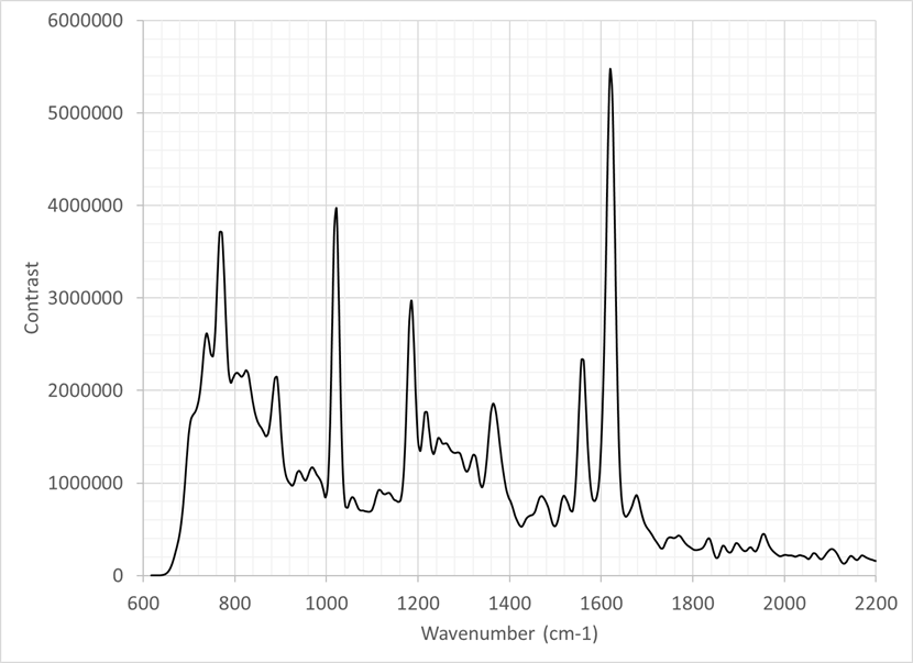
The figure displays the Raman spectrum acquired from an equine synovial fluid sample using ODIN. The data is acquired using a 30-second integration time and is the average of 10 frames. Limited processing has been applied to the data (e.g. removal of cosmic rays). The resulting power spectrum displays several discrete peaks observed from 750 cm-1 through to 1700 cm-1, of which several can be labelled based on known functional groups and amino acids. The results show ODIN could be used to study this and similar biological/biochemical materials non-destructively and without sample preparation constraints, as demanded by other techniques including SERS.


